Name: TEOH LEONG SIN
Matrix no: 111433
Lab 1: Principle and use of microscope
1.1 Setting up and using microscope
Introduction:
In order to be seen, microorganisms need to be magnified. Despite advances in other area of microscopy (for example, the electron microscopy), the light microscopy is still the instrument most frequently used for viewing microorganisms.
Objective:
Learn to use a simple bright-field microscope correctly.
Results:
Total magnification = objective lens power x eyepiece lens power(10x)
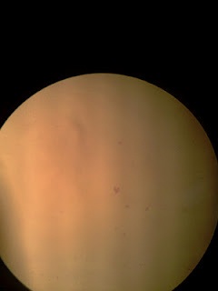
Salmonella enteritidis under 40x magnification:
Salmonella enteritidis under 100x magification:

Salmonella enteritidis under 400x magnification

Discussions:
1) Specimen is observed from lowest magnification to highest magnification (x40,x100,x400)
2) Light intensity is adjusted to observe the specimen clearly.
3)Specimen focused by adjusting fine and coarse adjustment knob to obtain a view of specimen.
4)Colony marphology of salmonella enteritidis:
a)Shape:circular
b)Size: tiny (punctiform)
c) Surface: shiny and smooth in appearence
d)Color: Red
Conclusion:
I could get a clearer image of Salmonella enteritidis in the magnification of x400. This mean the higher the magnification of objective lens, the clearer the view of image.
References:
http://en.wikipedia.org/wiki/Salmonella
http://quizlet.com/370451/microbiology-student-presentations-flash-cards/1.2 Examination of cells
Because of their extreme minuteness, bacteria are not generally studied with the low-power or high power-power dry objectives. Instead they are stained and observed with the oil immersion objective.
The wet mount methods enables you to study the sizes and shapes of living microorganisms (drying or staining microorganism distort them). It also enables you to determine if cells are motile. The wet mount method is quick and easy, and does not require special equipment.
Objective:
-To provide an experience in the use of microscope
- To illustrate the diversity of cells and microorganisms.
Saccharomyces cerivisiae (yeasts) under x1000 magnification:

Lactobacillus fermentum under x1000 magnification:

Discussion:
1)The oil immersion fills the space between the objective lens and specimen and matches the refractive index of the glass coverslip and glass objectives lens. At a given focal length, this allow me to achieve a greater numerical aperture.
2)There are 2 different types of microorganisms which been observed:
a)Lactobacillus fermentum
b)Saccharomyces cerivisiae
3)Colony marphology of Lactobacillus fermentum
-Shape:Rod
-Size:Tiny
-Surface:Smooth
-Texture:moist
-Color:Violet
4)Colony marphology of Saccharomyces cerivisiae
-Shape:Circular
-Size:Tiny
-Surface:Smooth
-Texture:Moist
-Color:Lemon yellow
Conclusion:
Oil immersion with 1000x magnification lens allow us to observe the specimen more detail which include shape,size,surface,texture and color.
References:
http://en.wikipedia.org/wiki/Saccharomyces_cerevisiae
http://onlinelibrary.wiley.com/doi/10.1046/j.1365-2958.2003.03332.x/full
http://microbewiki.kenyon.edu/index.php/Lactobacillus
http://en.wikipedia.org/wiki/Lactobacillus_fermentum


